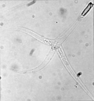
An animal in disguise
For over a century, scientists classified Myxozoa as single-celled protozoans. Their microscopic size and simple structure seemed to confirm this identity. However, genetic analysis in the 1990s revealed a startling truth: myxozoans are not protists but highly evolved animals. Specifically, they belong to the phylum Cnidaria, making their closest relatives jellyfish, sea anemones, and corals. This reclassification required a radical shift in understanding, showing a case of extreme evolutionary reduction.
Myxozoans descended from free-living, jellyfish-like ancestors but adapted to a parasitic lifestyle by shedding complexity. They lost genes responsible for development, cell-to-cell communication, and coordination. The result is one of the most structurally simple animals known, some consisting of only a handful of cells. They lack a mouth, a gut, and a nervous system. Their genomes are also among the smallest in the animal kingdom, evidence of their simple existence. One species, Kudoa iwatai, has a genome of just 22.5 megabases, with only about 5,500 protein-coding genes.
The parasite's toolkit
Despite their simplicity, myxozoans have an important feature linking them to their cnidarian kin: stinging cells. What were once called "polar capsules" are now recognized as nematocysts, the harpoon-like organelles that jellyfish use to capture prey. Myxozoans use these microscopic weapons not for predation, but to anchor themselves to a host to begin the infection process. The structure of these capsules, built from unique proteins like minicollagens, shows their shared ancestry with free-living cnidarians.
Most of the more than 2,180 described species of Myxozoa have a complex two-host lifecycle. This typically involves a vertebrate, usually a fish, and an invertebrate, such as an aquatic annelid worm. A well-known example is Myxobolus cerebralis, which causes "whirling disease" in salmon and trout. The parasite infects the cartilage of young fish, causing skeletal deformities and neurological damage. This damage often results in a characteristic corkscrew swimming pattern that gives the disease its name. The parasite's spores are released when the fish dies, consumed by tubifex worms, and develop into a new infective stage that is then released into the water to find the next fish host.